Clinical Evidence · Peer-Reviewed Research

Clinical Evidence & Research

Evidence base for DBD cold atmospheric plasma (CAP) technology in wound healing and biomedical applications — curated core publications with data-driven insights.

1
Randomized Controlled
Trial (RCT)
2
SCI Research
Papers
43
RCT Enrolled
Patients
62
RCT Enrolled
Wounds
✦ Randomized Controlled Trial
Effect of Cold Atmospheric Plasma Therapy vs Standard Therapy Placebo on Wound Healing in Patients With Diabetic Foot Ulcers: A Randomized Clinical Trial

JAMA Network Open (2020) · Diabetes and Endocrinology

Journal: JAMA Network Open (2020) · Section: Diabetes and Endocrinology

DOI: 10.1001/jamanetworkopen.2020.10411

Citation: Stratmann B, Costea TC, Nolte C, et al. Effect of Cold Atmospheric Plasma Therapy vs Standard Therapy Placebo on Wound Healing in Patients With Diabetic Foot Ulcers: A Randomized Clinical Trial. JAMA Network Open. 2020;3(7):e2010411.

Key Findings

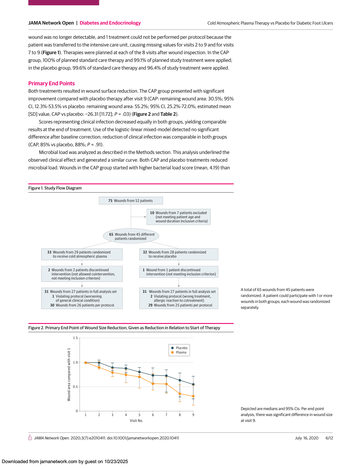
30.5% vs 55.2%
Residual wound area in CAP group vs placebo. CAP reduced wound area by nearly 70%.
P = 0.03 *
−26.31 relative units
Difference in wound area reduction between CAP and placebo (mixed-model estimated mean).
95% CI, P = 0.03
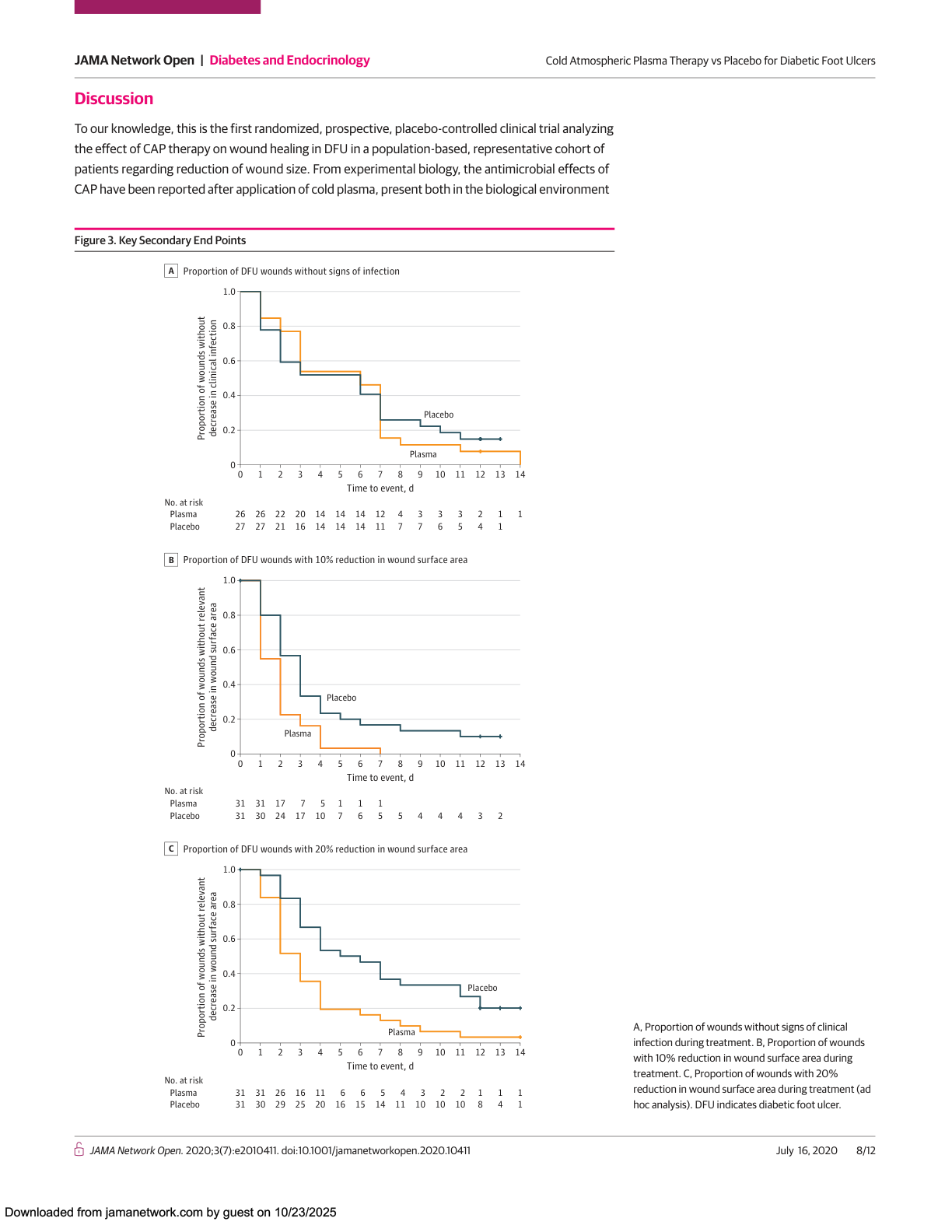
10% reduction — significantly earlier
All wounds in the CAP group achieved ≥10% reduction; 3 placebo wounds never did.
P = 0.01 *
20% reduction — significantly earlier
CAP group reached 20% wound area reduction significantly faster (accelerated failure time model).
P = 0.001 **

📊 Primary Endpoint: Wound Area Reduction

100% 75% 50% 25% 0%
30.5%
CAP
55.2%
Placebo
P = 0.03 *
Residual Wound Area After Treatment

CAP group residual 30.5% (95% CI: 12.3%–53.5%), placebo group residual 55.2% (95% CI: 25.2%–72.0%). Estimated mean difference −26.31 ± 11.72.

JAMA Figure 1+2: CONSORT flow and wound area reduction Original Figure 1 (CONSORT Flow Diagram) & Figure 2 (Wound Area Reduction)
📈 Secondary Endpoint: Time to Wound Reduction (Kaplan–Meier Curves)

Wound Area Reduction ≥10% 0 0.5 1.0 Proportion Not Reduced 0 2 4 7 10 Days CAP Group Placebo Group P = 0.01 Wound Area Reduction ≥20% 0 0.5 1.0 0 2 4 7 10 Days CAP Group Placebo Group P = 0.001 ** Time (Days)

Accelerated failure time model analysis. Median time to wound reduction was significantly shorter in the CAP group compared to placebo.

JAMA Figure 3: Kaplan-Meier curves Original Figure 3A–C: Kaplan–Meier Curves for Infection Resolution, 10% Reduction, and 20% Reduction

✦ Mechanistic Study

Nanosecond Pulsed DBD Plasma Generates Reactive Oxygen Species That Trigger Immunogenic Cell Death via Intracellular Oxidative Stress in A549 Lung Cancer Cells

International Journal of Molecular Sciences (2017)

Journal: International Journal of Molecular Sciences (2017) · Authors: Abraham Lin, Billy Truong, Sohil Patel, et al.

Submitted 2017-03-01 · Accepted 2017-04-28 · Published 2017-05-03

Citation: Lin A, Truong B, Patel S, et al. Nanosecond Pulsed DBD Plasma-Generated Reactive Oxygen Species Trigger Immunogenic Cell Death in A549 Lung Carcinoma Cells through Intracellular Oxidative Stress. Int J Mol Sci. 2017;18(5):966.

Key Findings

68×
ATP secretion surged from 12.3 nM to 831.2 nM after DBD plasma treatment.
"Find me" danger signal
4h
Intracellular ROS peaked 4 hours after plasma treatment.
Not restored to baseline at 24h
300 mJ
Optimal energy parameter — ~50% cell viability with strongest immunogenic death signaling.
Dose-dependent effect
↑ Macrophages
Conditioned medium from plasma-treated cancer cells enhanced macrophage phagocytosis and killing activity.
TNF-α / IL-6 elevated

📊 ATP Secretion (Immunogenic Cell Death Marker)

0 200 400 800 Extracellular ATP Concentration (nM) Control 12.3 nM DBD Plasma 831.2 nM 68× Increase

Measured 10 minutes post-plasma treatment. ATP is a key "find me" signal for immunogenic cell death.

📈 Intracellular ROS Time Course

0 50 100 0h 1h 4h 24h Time After Plasma Treatment Baseline Peak Not recovered NAC (antioxidant) reverses

ROS-positive cell proportion peaked at 4 hours post-DBD plasma treatment and had not returned to baseline by 24 hours.

🔬
Core Mechanism

DBD plasma-generated reactive oxygen species (ROS) trigger immunogenic cell death (ICD) via the intracellular oxidative stress pathway, releasing "danger signals" that activate the immune system.

🧪
Key Evidence

① Intracellular ROS elevation
② CRT surface exposure on cells
③ Massive ATP secretion (68×)
④ Enhanced macrophage anti-tumor activity

💡
Clinical Significance

Demonstrates that DBD plasma is not merely a physical sterilization tool — it can exert therapeutic effects through immune mechanisms, providing a theoretical basis for plasma in onco-immunotherapy.

✦ Veterinary Application Review

Cold Atmospheric Plasma for Wound Healing in Veterinary Medicine — A Review of Preclinical Evidence

Evidence summary based on multiple animal studies and companion animal clinical cases

Source: Literature review · Evidence synthesis from animal experiments and veterinary clinical cases

Animal Models & Key Evidence

🐭
Murine model: Full-thickness wounds treated with plasma showed accelerated healing within 1 week, with increased angiogenesis and reduced scar formation.
🩺
Diabetic animal model: Plasma modulated the inflammatory response and activated repair cells, outperforming conventional pharmaceutical treatments.
🐑
Large animal models (sheep/pig): Significant bactericidal effect, promoted granulation tissue growth, and reduced infectious complications.
🐱🐶
Companion animal clinical cases: 7 cats and dogs with chronic non-healing wounds (up to 80 months duration) achieved complete healing after plasma therapy. Drug-resistant fungal infections also resolved.
🧹
Antimicrobial Action

Directly kills wound bacteria (including drug-resistant strains), reducing infection burden.

🔧
Tissue Repair

Reactive species activate Nrf2 and other cell signaling pathways, promoting epidermal cell migration and collagen deposition.

🌡️
Anti-Inflammatory Modulation

Suppresses excessive inflammation while recruiting immune cells to clear necrotic tissue.

✦ Clinical Study Protocol

Cold Plasma Therapy for Acceleration of Wound Healing in Diabetic Foot — Study Protocol KPW2016-1.1

Cold Plasma Therapy for Acceleration of Wound Healing in Diabetic Foot — Study Protocol KPW2016-1.1

Protocol ID: KPW2016-1.1 · Principal Investigator: Prof. Dr. Dr. D. Tschöpe, Herz- und Diabeteszentrum NRW, Ruhr-University Bochum

Protocol Overview: Prospective, randomized, patient-blinded, placebo-controlled clinical trial. Conducted at 2 German clinical centers (Herz- und Diabeteszentrum NRW + Klinikum Karlsburg). Ethics approval: Ruhr-University Bochum Ethics Committee. ClinicalTrials.gov ID: NCT04205942.
🎯
Study Design

Randomized, patient-blinded, placebo-controlled. Patients are masked to treatment arm (device sound mimics placebo).

📋
Treatment Regimen

Once daily for 5 consecutive days → every other day for 3 sessions, totaling 8 treatments within 14 days. CAP 30 s/cm² delivered via sterile spacer at optimal distance.

📊
Primary Endpoints

① Wound area reduction ② Clinical infection improvement ③ Microbial load reduction. Secondary: time to reduction, quality of life, etc.

WhatsApp
WeChat QR Code
WeChat Customer Service
Michael.Tsai
Scan to add friend and inquire about products